What is NANOFAT TRANSFER FOR ACNE SCAR
What is NANOFAT TRANSFER? Acne scars are usually the result of inflamed blemishes caused by skin pores engorged with excess oil, dead skin cells and bacteria. The pore swells, causing a break in the follicle wall. Shallow lesions are usually minor and heal quickly. But if there is a deep break in the wall of the pore, infected material can spill out into surrounding tissue, creating deeper lesions. The skin attempts to repair these lesions by forming new collagen fibers. These repairs usually aren’t as smooth and flawless as the original skin.
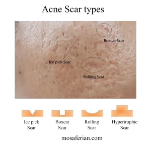
There are four types of acne scars:
1. Rolling scars: Broad depressions with sloping edge.
2. Boxcar scars: Broad depressions with sharply defined edges.
3. Atrophic scars: Flat, thin scars or depressed scars.
4. “Ice-pick” scars: Usually small, yet obvious holes in the skin.
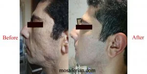
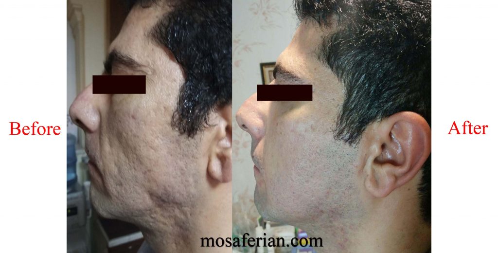
Nanofat grating for acne scar written by Dr. Mosaferian
https://www.imcas.com/es/academy/blog/795/nanofat-transfer-for-acne-scar
Nanofat Transfer
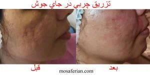
Nanofat Transfer refers to the very small particles of fat that are specifically filtered and emulsified and used for areas to be injected, in this process the harvested fat filtered and emulsified to obtain nano-particles.
This emulsion contains high percentage of stem cells, endothelial cells, monocytes, macrophages and granulocytes which activation of stem cells increases collagen production. During processing and filtering of harvested materials, many fat cells die (cell apoptosis).
Nanofat grafting has been shown to have beneficial effects in the treatment of scars, hyperpigmentation, and dark spots. However atrophic and rolling scars as well as hyperpigmentation had greatest improvement with this treatment. In our practice sharp needle intradermal fat grafting (SNIF) had greatest response for the treatment of atrophic and rolling scars and topical application of nanofat after skin resurfacing techniques would improve dark spots as well as post inflammatory hyperpigmentation (PIH).
Effects of nanofat on PIH
Treatment of post inflammatory hyperpigmentation (PIH) with best topical and systemic approaches last for several weeks and may even last for more than 5 years if left untreated. This type of pigment is different than normal dark spots because it is a result of inflammation. PIH with other causes such as complication of energy-based devices would aggravate with further treatments and manipulations, however nanofat application in combination with traditional treatments had rapid and significant improvement in such cases.
The most important step of all is to protect treatment areas from the sun. UV rays are the enemy of acne scars, dark spots and pigmented areas. They trigger inflammation and spike melanin production, causing dark spots and PIH to become even darker and harder to fade.
Recovery
To minimize swelling in the face one may use cool, clean compresses or ice wrapped in a dry cloth. Mild cooling of the area will help reduce inflammation and rapid recovery. however, too much ice may limit blood circulation to the area and conversely increase inflammation and pigmentation.
How can you tell the difference between a seroma n fluid filled tunnel after liposuction? Should a seroma or fluid filled tunnel be treated after liposuction, if so how long after liposuction n how should it be treated?
Thank you for your question
Fluid accumulation after surgery is called a seroma.
If it fluid collection forms i.e. the patient develops a seroma then this should be treated correctly.
Fluid collections should be drained on a regular basis until it no longer forms. Preferably this means being seen on a daily basis draining the fluid until it no longer accumulates.
Failure to appropriately manage a fluid buildup can lead to an encapsulation of the seroma, causing a chronic seroma to form.
Once the seroma becomes chronic or encapsulated, it can no longer be treated by just draining the fluid on a regular basis. An encapsulated seroma needs to be excised with open surgery.
Most fluid collections can be managed and eliminated by being drained on a regular basis over a few weeks after the procedure. Failure to manage seromas correctly can lead to chronic seroma formation, which is far more difficult to manage and can leave secondary negative consequences, such as unnecessary, incisions and scars.
Best of lucks