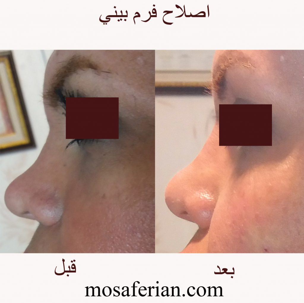
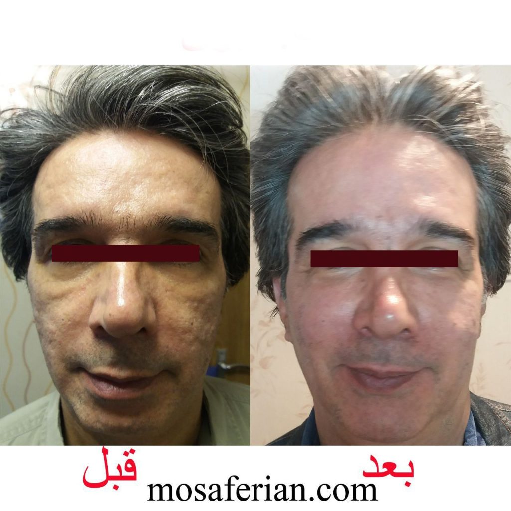
Category: Filler injection

  • Radiesse filler

    Radiesse filler

    Radiesse and Radiesse(+) comprise the first and only CaHA portfolio available that stimulates collagen and elastin production for both immediate, natural-looking results and long-term improvement. The microspheres then dissolve naturally through the normal metabolic process. 

    radiesse filler

    Radiesse smooths lines and adds volume in the mid and lower face. Calcium hydroxylapatite (CaHA) is what makes Radiesse and Radiesse (+) unique injectable aesthetic treatment options. These calcium-based microspheres are similar to minerals that are found naturally in your body.

    • Offers immediate improvement you want, the collagen and elastin you need.
    • Has long-lasting results: 12+ months or more in most patients4,7
    • Is the first and only CaHA injectable treatment in Canada
    • Has a well-established safety profile and predictable results

    How long does the effect of CaHA last?

    • Results are long lasting, but not permanent. How long the effect of treatment lasts is dependent on the individual. There are many factors to consider, such as the structure of the skin, your lifestyle and age. Studies show that in nasolabial fold treatments (the lines that run from the nose to the corner of the mouth), for example, results last up to 12 months. The procedure can be repeated, please consult with your healthcare professional about what is appropriate for you.

    Can Radiesse be combined with other products?

    For a holistic treatment, your healthcare professional may advise combining RADIESSE® with hyaluronic acid dermal fillers such as BELOTERO®, or other treatment options such as Ultherapy and NEOCUTIS®*. Please consult with your healthcare professional

    radiesse filler

    Radiesse for Men

    Male skin aging is a gradual, ongoing process that is strongly affected by environmental factors. Sun exposure, poor nutrition, and occupational or personal stress take their toll on male skin, especially on the skin’s main structural competent – collagen. The consequences: firm skin loses its elasticity and wrinkles and folds begin to form. Fortunately, men can address these concerns by choosing a treatment strategy with Radiesse, which leads to natural looking results without compromising masculinity. RADIESSE is the only dermal filler that works in such a way that it can immediately reduce facial wrinkles and continue to provide structure over time, with results that last a year or more in many patients.

    Source

    https://www.merzaesthetics.ca/solutions/radiesse/

  • Volumetric Effect and Patient Satisfaction after Facial Fat Grafting

    Volumetric Effect and Patient Satisfaction after Facial Fat Grafting

    Background: 

    Facial fat graft decreases in volume after transplantation. This observation is based on overall facial three-dimensional analyses, because there is sparse information on volume changes in well-defined aesthetic areas. The authors aimed to assess the overall and, more specifically, the local volumetric effects of facial fat grafting and relate these effects to patient satisfaction up to 1 year after treatment.

    Methods: 

    All consecutive adult female patients who were scheduled for facial fat grafting without additional surgical procedures were asked to participate. All patients underwent the same fat grafting method. An algorithm-based personalized aesthetic template was applied to define specific aesthetic areas on the preoperative three-dimensional image. Objective outcome parameters [i.e., three-dimensional volume differences, patient satisfaction (FACE-Q questionnaire)] were measured at baseline and at 6 weeks, 6 months, and 12 months after fat grafting.

    fat graft to face

    Facial fat graft results: 

    Of 33 female patients who underwent a facial fat graft procedure, 23 patients had complete three-dimensional data and were eligible for analysis. The highest volume gain was observed 6 weeks after grafting and was followed by a gradual loss thereafter. Overall and in the zygomatic area, a substantial gain in volume was still present 1 year after grafting, whereas this effect was lost in the lip area. FACE-Q scales Satisfaction with Facial Appearance Overall and Satisfaction with Cheeks improved too, whereas scores for Lines: Lips returned to baseline levels. The improvement in FACE-Q scales was in agreement with the objective change in volume.

    Conclusion: 

    Gain in overall and local volumetric effects is accompanied by comparable changes in patient satisfaction.

    CLINICAL QUESTION/LEVEL OF EVIDENCE: 

    Therapeutic, IV.

    Reference:

    https://journals.lww.com/plasreconsurg/abstract/2022/08000/volumetric_effect_and_patient_satisfaction_after.13.aspx

  • Under Eye Filler Before And After Photos❤

    Under Eye Filler Before And After Photos❤

    What is under eye filler? A tear trough is a deep crease between the lower eyelid and upper cheek, and as we age we can experience volume loss in this area which can leave dark circles, shadows and hollows around the delicate under-eye area. This can often make us look tired, older, haggard and run down, even when we are perfectly rested and well hydrated. This can be easily treated with under eye filler.

     cheek augmentation & under eye filler before and after
    cheek augmentation &tear trought correction

    Under eye filler bruising

    One of the most common side effects of dermal fillers is bruising. Even if you have never bruised before, it is always a possibility. Why? Because bruises are just a small hematoma; they occur when small blood vessels get punctured and leak into the soft tissue beneath. The good news is there are some tips and tricks up our sleeve that you can use to help reduce the bruising pre- and post-treatment.

    At  Niayesh Beauty Clinic we use FDA- approved dermal filler and Cannulas are used to place filler under the very thin muscle layer, which results in a more natural appearance with minimal-to-no bruising.

    Under eye filler before and after

    Pre-Appointment Tips

    • There are medications that you may want to consider stopping 1 week prior to your appointment (and with your doctor’s consent.) These medications thin your blood, making it more difficult for it to clot, increasing the likelihood you will bruise following your procedure. This list includes:
      • Aspirin
      • Excedrin
      • Ibuprofen (Motrin, Aleve, Advil)
      • St. John’s Wart
      • Fish Oil
      • Vitamin E
      • Ginko Biloba
      • Ginseng
      • Fenugreek
      • Garlic
    • No alcohol or green teas 1-2 days before injections
    • Eat fresh pineapple, it contains bromelain which has been know to decrease bruising
    • Take Arnica tablets for a couple of days leading up to your appointment. Arnica is a homeopathic remedy to help prevent bruising/swelling.
    • under eye filler

    tear trough correction before and after
    under eye filler

    Tear trough filler aftercare

    Congrats on your new fillers! Here are a few tips to avoid bruising after your procedure:

    • Avoid alcohol for 1-2 days after injections
    • Avoid vigorous exercise for 1-2 days
    • Wait 24 hours before applying makeup
    • Apply Arnica gel, it will help you heal faster
    • Apply ice packs. The ice contracts the blood vessels and lessens the chance that your bruise will spread.

    While these tips and tricks can help, we always recommend planning to get your fillers done 1-2 weeks before any big event or photoshoot. It is best to aim for 2 weeks if you are someone who bruises easily.

    tear trought correction
    tear trought correction

    Recovery time

    Dermal fillers work very quickly, and you won’t have to wait twelve months to see the full benefits of your injections. That said, these injectable treatments take some time to integrate into your tissues, and it’s normal for your dermal filler to take up to two weeks to fully settle into your face.

  • Russian Lip Filler Before And After💋

    Russian Lip Filler Before And After💋

    What is russian lip filler? This new trend has introduced a brand new shape to the lips, providing a flatter, wider look to match the one supported naturally by Russian women. The trend began due to the desire for lips that are larger in volume and plumpness, without the worry of filler migration or the ‘duck lips’ and ‘trout pout’ look that many want to avoid when having lip filler. Russian Lips derive from the ‘Russian Doll’, where they form a heart shape in the centre with a flatter, higher appearance.

    However this look isn’t for everyone, and although this technique does avoid filler migration, it doesn’t necessarily look more natural than the more common lip filler technique.

    russian lip filler before and after
    russian lips

    Russian lip filler technique

    The technique involves injecting tiny droplets of dermal filler into the lip, mainly focused around the centre to achieve the heart-shaped look. Filler is injected vertically into multiple injection points and dragged upward from the base to the top of the lip, rather than horizontally with traditional lip augmentation. Russian Lips are larger in height rather than volume, which requires a longer treatment time due to an increase of product needed to achieve the desired look.

    lip augmentation before and after
    russian lips

    Russian vs French Technique

    So what is the big difference between standard lip filler (French Technique) and Russian lip filler technique?

    Typically, French lip fillers are injected horizontally into the lip, resulting in an evenly spread volume and fullness effect. However, in order to execute the Russian filler technique, your provider will opt for a smaller syringe and inject small amounts of filler vertically, focusing in on the center of the lips. Another key difference is that the Russian lip filler technique aims to heighten the lip, focused in on the center, rather than to add overall plumpness throughout the lips, thus providing that heart-shaped look. 

    Additionally, your Russian treatment may take longer than a standard lip filler appointment due to the necessary precision required to fill only a specific region of the lips. The procedure may last approximately between thirty minutes to an hour, and may result in additional bruising and swelling , which is completely normal and temporary. Your provider will also be able to relay an accurate timeline for the lifespan of your Russian lips, but generally your Russian lip fillers will last as long as standard lip fillers do, being anywhere from six to twelve months.

    lip filler french style
    French Lip Filler

    Side Effects

    After receiving lip filler injections, swelling can be easily managed by applying ice to the lip area. Gently apply an ice pack wrapped in a towel or a cold compress to the lips. Be sure to use soft cloth and do not to use anything that could stick to the lips, as this could worsen the discomfort.

    It is recommended that you drink plenty of water and eat lots of healthy fruits and vegetables after your procedure. Avoid very salty or spicy foods, as these can irritate the lips. Staying hydrated and eating well can help the body heal.

    At night, you may wish to sleep with your head elevated to reduce blood flow and fluid retention in the area overnight. Avoid sleeping on your face.

    Having said that, swelling should go down within 2–3 days after your lip filler treatment, and should subside completely within 2 weeks post-treatment.

    Russian Lip Filler Cost

    How Much Do Russian Lips Cost? Just like any dermal filler application, you pay for the cost of each syringe used in the process. So, the price for the filler is no different for Russian lips, however the technique often requires more filler to be used.

    lip filler
  • Arnica Cream For Bruises In 2024🌻

    Arnica Cream For Bruises In 2024🌻

    Arnica cream seems to have a mitigating effect on ecchymosis, most notably following rhinoplasty and facelifts/facial procedures. Bromelain is well supported across numerous studies in reducing trismus, pain, and swelling following molar extractions. However, there was no effect demonstrated when evaluating topical arnica following blepharoplasty procedures. Arnica cream known to have anti-inflammatory, antimicrobial, and analgesic properties.

    Arnica Montana, the scientific name of the Arnica plant, has been used widely for over centuries to  heal bruises , wounds and reduce pain and swelling.

    A systematic review of the literature demonstrates the potential for arnica cream to improve perioperative outcomes including edema, ecchymosis, and pain control.

    Arnica flower

    Arnica cream for bruises

    Arnica can be used to decrease swelling, bleeding, bruising and reduce pain. Arnica creams  can be used for topical application to heal bruises  after filler injection and subcision effectively.

    Using the topical arnica  two to three times a day for three weeks is suggested to get the desired results. With consistent use, people have found it to reduce pain, bruising, stiffness, and swelling.

    Visit on Amazon

    Arnica  side effects

    Arnica is considered unsafe for ingestion by the FDA. Consuming arnica can lead to:

    • diarrhea
    • vomiting
    • nausea
    • internal bleeding

    How to use Arnica cream?

    Arnica is a safe and effective option to deal with bruises and pain. Make sure not to apply arnica cream to any open areas of skin and rather let them start to heal up before starting to apply arnica to deal with the surrounding bruising.

    As with any skin care product, discontinue use if irritation, redness, swelling, or hive-like rash occur. Allergic contact dermatitis to any skin product can occur but it is rare.

     How long does it take for Arnica cream to work on bruises?

    After using arnica for a total of 4 days the bruises were barely noticeable. Without the cream, bruises that dark would have taken about 7 to 10 days to completely heal.

    Why can bruising occure after Botox or fillers?

    Bruising doesn’t always occur after Botox or dermal fillers, but any time an injection is made under the skin, bruising is possible. Black, red or purple bruises form when damaged blood vessels leak blood into the surrounding tissue. This can occur if a blood vessel is nicked by a needle, or if skin is pinched or pulled, causing capillary damage.

    Arnica cream

    Who is most likely to bruise after Botox or fillers?

    Genetics play a role in likelihood of bruising. Women are more likely to bruise than men, and older people are more likely to bruise than those who are younger. In addition, the following avoidable factors may increase your risk of bruising:

    • Use of blood thinners, including aspirin and NSAIDs
    • Frequent use of alcohol, which increases blood flow
    • Exercising immediately before or after treatment
    • Systemic inflammation due to poor diet or poor health

    Bruise prevention after Botox or filler

    After your filler or Botox treatment consider taking Arnica  cream, a natural remedy with the power to reduce post-procedure pain and treat bruising. Arnica Montana can either be taken in pill form or topically applied to the treatment area. Some authors may even recommend taking the supplement a few days before your injectable treatment to further prevent bruising.

    Arnica cream

    Arnica
  • Hyaluronidase protocol for dermal fillers

    Hyaluronidase protocol for dermal fillers

    New High Dose Pulsed Hyaluronidase Protocol for Hyaluronic Acid Filler Vascular Adverse Events

    Claudio DeLorenzi, MD, FRCS

    The purpose of this article is to update the changes to the author’s protocols used to manage acute filler related vascular events from those previously published in this journal. For lack of a better term, this new protocol has been called the High Dose Pulsed Hyaluronidase protocol(HDPH) for vascular embolic events with hyaluronic acid (HA) fillers. The initial protocol used involved many different modalities of treatment. The current protocol is exceedingly simple and involves solely the use of hyaluronidase in repeated high doses. Despite the simplicity of the treatment, it has proven itself to be very successful over the past two years of clinical use.

    There has been no partial or complete skin loss associated with this protocol since its implementation if the protocol was implemented within 2 days of the ischemic event onset. The protocol involves diagnosis and repeated administration of relatively high doses hyaluronidase (HYAL) into the ischemic tissue repeated hourly until resolution (as detected clinically through capillary refill, skin color, and absence of pain). The dosage of HYAL varies as the amount of ischemic tissue, consistent with the new underlying hypothesis that we must flood the occluded vessels with a sufficient concentration of HYAL for a sufficient period of time in order to dissolve the HA obstruction to the point where the products of hydrolysis can pass through the capillary beds.

    Although vascular embolic events are rare, it is important to note that the face has higher risk and lower risk areas for filler treatment, but there are no “zero risk” areas with respect to filler treatments. Even with good anatomic knowledge and correct technique, there is still some nonzero risk of vascular embolic events (including highly skilled, experienced injectors). However, with careful low pressure, low volume injection technique, and adequate preparation for treatment of acute vascular events, the risk is quite manageable and the vast majority of adverse events are very treatable with an excellent prognosis, with a few exceptions. This new protocol offers excellent results, but requires further research to determine optimal parameters for various HA fillers.

    Hyaluronidase Protocol:

    ESSENTIAL CONCEPTS REGARDING RESOLUTION OF FILLER-RELATED VASCULAR ADVERSE EVENTS

    This article summarizes a new approach to the management of acute accidental intravascular embolism with hyaluronic acid (HA) fillers. As with the former protocol previously published,1 avoidance of complications is still the best strategy (Table 1). The old protocol involved a daily single treatment with hyaluronidase (HYAL) of 450 to 600 iu, as well as other modalities of treatment such as nitropaste, hyperbaric oxygen etc. In contrast, the new  hyaluronidase protocol involves HYAL dosing based on the volume of ischemic tissue, with hourly repeated dosing to maintain high concentrations of HYAL throughout the ischemic zone. Treatment is based on the clinical determination of the approximate surface area of ischemic tissue (as determined clinically by observation of skin color and capillary refill), with larger doses of HYAL used for larger areas of involvement. This report is based on the author’s clinical experience as a consulting physician with several dozen cases in the past two years using this new high dose hyaluronidase protocol, in comparison to clinical experience in over 50 cases with previous hyaluronidase protocols.

    The author assumes that the pathology of filler related ischemia is due to arterial embolism of filler that typically occurs immediately at the time of filler treatment, and that the solution is to flood the tissues containing the obstructed vessels with HYAL in sufficient concentration for a sufficient length of time to relieve the HA obstruction. Rarely, the clinical history is that the patient was completely normal at the termination of treatment, and that signs of ischemia started at some time (typically hours) later. This phenomenon of “delayed onset” is discussed further below.

    The natural history of vascular embolic events (ie, untreated) progresses from momentary blanching (which may only last a few seconds), through livedo reticularis (up to a few days), to blisters (day 3), crusting, necrosis, slough, and finally healing by secondary intention − a process that may take six weeks or more. If untreated, it takes approximately 3 days before blisters appear on the skin (this seems to be a relatively consistent finding), and frank necrosis may not be evident for several days thereafter (typically after day 6), but the signs of ischemia are generally present right from the very beginning if you look for them. With appropriate treatment with the new High Dose Pulsed Hyaluronidase protocol, we typically see complete reversal of all the signs of ischemia and complete return to normal.

    Thus, instead of 6 weeks or more of slow healing by secondary intention, we see complete resolution with no signs of secondary problems within 3 days of the event, with patients typically suffering no more than a few bruises and so called “injection site reactions” (the normal sequelae of repeated needle injections). Diagnosis is completely clinical in nature. It involves examination of the skin, noting its color and in particular its capillary refill time. Typical cases of vascular obstruction may show some blanching, but this is often missed, since it is only momentary. A mottled skin appearance, termed livedo reticularis, is almost always apparent (except in cases of severe bruising).

    Skin capillary refill test

     Capillary refill time is noted to be very slow. Using the finger holes of an instrument (such as the non business end of a pair of suture scissors, for example) help to assess the capillary refill (Figure 1).(A) When determining the status of the skin’s capillary refill time, it is helpful to use and instrument to compress the skin. Patterns that are not typical in nature are more easily discernible. (B) The objective of this clinical test is to compare the refill time of the zone in question to normal skin either adjacent or on the contralateral side.

    The first major breakthrough in the treatment of vascular adverse events (AE), was the administration of HYAL (the author believes that the ancillary treatments such as nitropaste etc. probably did not contribute very much to the final outcome). With the old hyaluronidase protocol, the author occasionally saw some blistering, crusting, and eventually some mild scarring, mild to moderate dyschromia (particularly in patients of color) despite treatment with HYAL. These occasional problems seemed particularly worse if more regions of the face had been originally involved in the vascular AE (eg, ischemia of the ipsilateral upper and lower lip, nose, and glabella) compared to single site (lip ischemia alone).

    This suggested to the author that perhaps we were not using enough HYAL in at least some of these patients (otherwise, why did some patients heal completely, while others had crusting and scars, dyschromia, etc?). This lead to the “HYAL flooding hypothesis” (further discussed below), namely that HA filler obstructed arteries needed to be bathed in high concentrations of HYAL for long enough periods of time to dissolve the filler. This concept is illustrated in Figure 2, showing that the recommended dosage of HYAL increases stepwise with the numbers of anatomical areas involved. If more parts of the face are involved with ischemia, then larger doses of HYAL will be needed to treat that individual. We use ischemic surface area as a proxy for tissue volume (not the volume of intravascular filler − the amount of filler within the intravascular space is essentially irrelevant, and not clinically observable at the bedside). We can assume that all the arteries in the ischemic areas of the face are completely filled with HA filler. In order to obtain a sufficient concentration of HYAL (ie, mg/cc3) a larger volume of tissue (the denominator) means that a larger amount of HYAL is required.

    Some common patterns of injury, and the typical doses used by the author for each. HYAL is injected into the ischemic tissues and gentle massage is then used to maximize the contact time between the ischemic tissues and HYAL. The tissues may be edematous, but gentle massage following injection of HYAL will tend to decrease this. Massage is important to help maximize contact between hyaluronidase and the filler causing the obstruction.

    before & after vascular occlusion treated with hyaluronidase
    before & after vascular occlusion treated with hyaluronidase

    Analysis of Hyaluronidase Protocol

    The newest hyaluronidase protocol for the treatment of filler embolism involves several important changes. First came the realization that HYAL was the critical factor in treatment. None of the ancillary treatments previously recommended (topical nitropaste, hyperbaric oxygen, etc.) are used at all any more, except for one baby aspirin per day for seven days (ASA is given to reduce platelet activity, but direct evidence for its benefit is lacking in this situation). The author has not recommended or used any of the ancillary treatments for over 2 years now.

    With experience came the second important observation that dosing should depend on the quantity of tissues adversely affected (ie, one dose does not fit all). This is based upon the assumption that HYAL has to be used in quantities sufficient to bathe the obstructed vessels in a concentration sufficient to hydrolyse the filler causing the obstruction. A vessel that can no longer transport fresh blood because it is blocked by HA must be bathed (flooded) by a sufficient concentration of HYAL to diffuse across the arterial wall and then break down the HA to metabolic products small enough to pass through the capillary system. We cannot possibly know where the obstructed vessels are within the block of ischemic tissue. We must assume that all the vessels within the zone of ischemia are obstructed, and treatment must involve flooding the entire zone with sufficient HYAL concentration to promote complete hydrolysis of the HA filler (which, again, could be anywhere within the ischemic zone’s arterial system). With this view, partial breakdown of HA is insufficient, because partial breakdown products can still obstruct blood flow (although they may be pushed further downstream by arterial pressure). Thus when we are advising “HYAL flooding” what we mean to say is that we want to ensure that the obstructed vessels are soaking in a sufficient concentration of the enzyme to promote complete hydrolysis of the HA within the vessels. We assume that partial hydrolysis is insufficient. The author also suggests that partial breakdown may result in further obstruction downstream.

    In fact, in this view, partial breakdown may result in new areas of ischemia (or at least, a zone of injury of a different shape than at presentation, as is sometimes seen), as these breakdown products may be carried downstream past areas where collateral vessels are bypassing the initial obstruction. In fact, this is the mechanism by which we posit that the phenomenon of “delayed onset” ischemia occurs, when dilution or blood pressure moves an embolus further downstream. A vessel containing an embolus of filler might not be yet causing ischemia, since collaterals may initially be completely by-passing the obstruction. As the filler makes its way downstream, it may then block the collaterals and then manifest the ischemia.

    Clinical assessment of the patient must be ongoing and persistent. The goal of treatment is complete dissolution of the offending filler obstruction. To accomplish this, there must be sufficient concentration of HYAL in the right location long enough to result in (sufficiently) complete hydrolysis (Figure 3). The number of qualifications specified here are intentional, since a deficiency in any one of these elements is a recipe for failure to achieve the stated goal of complete resolution of the obstruction. In vitro, the response time for hydrolysis depends on the actual filler being tested. Let’s assume that a filler will take, roughly speaking, a few hours to break down, since the HYAL has to diffuse across the intact arterial wall and hydrolyse the filler. (Crosslinked fillers will take longer times to degrade with HYAL, whereas in contrast, in the author’s in vitro testing, non-crosslinked HA was hydrolyzed almost instantaneously, with complete liquefaction within just a few seconds of direct contact). It is known that HYAL itself is actively metabolized by the human body, so it is being deactivated at some rate as soon as it is injected. As swelling fluid accumulates from leaky capillaries in the ischemic environment, the HYAL is also being diluted. Finally, we know that as HYAL degrades the ground substance, it begins to diffuse away from the original injection site. Thus we can see that the amount of HYAL we originally injected into a region will be partially deactivated (by natural anti-HYAL agents), diluted by swelling fluids, and will physically diffuse away from the region where we want to maintain a high concentration. These three factors all act to reduce HYAL action, and the author suggests that these are reasons that we should act to top up the HYAL on a frequent basis to maintain the desired high concentration. The frequency of this topping up has to be determined in laboratory studies, but for now, the author has been using an hourly dosing schedule. This seems clinically to be safe and effective, but it may be overdoing it and dose ranging studies in a validated model are needed.

    duck lips before and after hyaluronidase
    Duck Lips

    INTRA-ARTERIAL VERSUS EXTERNALLY APPLIED HYALURONIDASE

    High volume vascular embolic events involve large bolus injections into the arterial system, and these typically require more vigorous treatment with Hyaluronidase because the zone of ischemia is larger, and dissolving the embolus takes longer. There is also the question regarding intra-arterial injection of HYAL, as opposed to the current strategy of simply bathing the external surface of the obstructed vessels with HYAL. With an arterial obstruction, as when clot busting drugs are used in intra-arterial treatments, there is the problem of maintaining the concentration of the drug where it is most needed. If the obstruction is complete, there is no flow past or through the obstruction, thus injecting HYAL into an occluded artery causes the HYAL to flow proximal to the obstruction. HYAL does work on the obstruction, but only at the site where it is exposed in the lumen of the vessel. In contrast, when flooding the entire zone of obstruction, the HYAL seems to break down the HA filler all along the vessel pathway, affecting the entire embolus at once. This hypothesis has not been tested however, and it remains to be proven whether external application of HYAL to the outside of a vessel is more or less effective than intra-arterial application of HYAL. Certainly for ease of use, external application of HYAL is very simple and appears to be safe and effective treatment, but there have been sporadic case reports at international meetings of treatment failures reversed with intra-arterial application of HYAL. This involves either a cut down procedure (to cannulate an artery) or super-selective angiography procedures that are not typically available except in tertiary care centers. For the time being, until evidence shows otherwise, external application by simply injecting HYAL into the affected areas and flooding the affected vessels through indirect diffusion appears to be a safe and effective solution in most cases. Clinical experience over that past few years has shown that the use of indirect soft tissue injection of HYAL alone without any ancillary treatments (ie, no nitropaste or hyperbaric oxygen etc.) has provided excellent results, superior to the previous protocol1 recommended.

    TIME OF ONSET

    Embolism probably most often happens contemporaneously with filler treatment, except in rare cases. There are some cases that report no evidence of problems at the end of treatment, but obvious signs of ischemia a few hours later. The author believes delay of onset of ischemia is rare, and at least some cases are likely due to perceptual blindness − ie, the injector simply is concentrating on the goals of symmetry, aesthetics, balance, and so on, and missing the signs of vascular ischemia.2 On occasion though, the author has heard of cases where the practitioner is adamant that the patient was completely fine at the end of treatment (ie, when the physician was actively assessing the patient for ischemia), only to discover rather obvious ischemia a few hours later. In these uncommon situations, the author hypothesizes that filler may be trapped in a vessel, perhaps at a bifurcation or branch point, with NO signs of distal ischemia because collateral vessels bypass the obstruction. At some point, this globule of filler may become dislodged by whatever mechanism, and then pass downstream to the precapillary arterioles, thus causing tissue ischemia. Despite the obvious violation of Occam’s Razor (where the simplest solution is usually correct), this two step process may turn out to have some validity in rare cases, but once again this is purely conjecture and the author has absolutely no hard evidence. In the typical event, the simple truth appears to be that the material seems to flow downstream within the arteries until it is constrained by size and can pass no further. It then simply blocks one or more arterial blood vessels, recalling that vessels can back fill towards a branch point by retrograde flow, and the filler can thus also obstruct nearby branches or even be passed towards a more distal zone through a main branch of a vessel. The zone of ischemia may be large or small sized depending on the amount of intravascular filler injected, and the relative importance of the specific blood vessel to the tissues in question. One could think of the vascular system as analogous to an Interstate highway system, where all points are connected, and it is quite possible to reach a distant exit ramp from any particular on ramp.

    MECHANISM OF INJECTION:

    HOW DOES FILLER GET INTO THE ARTERY?

    It may be that most of these events are a result of direct intra-arterial injection, with the opening of the needle or cannula directly within the lumen of the vessel. Clearly, aspiration before injection might provide good feedback if bright red blood is seen, but a negative result may be misleading.3 In areas of scarred tissues, as from previous trauma, or even from a long series of former filler treatments, some special considerations may come into play. As a needle or cannula is passed into the tissues, this may create an artificial pathway for the filler to flow. In fact, this is a technique used called pre-tunneling, where a needle or cannula is passed through the tissues, and the filler is slowly injected upon withdrawal. If the tissues are scarred, then the needle track acts as a conduit for filler to flow through (Figure 5). A possible mechanism for embolism becomes apparent. If a vessel is penetrated by a needle or cannula, even if the filler is deposited at some distance beyond the vessel, the filler may back track along the needle pathway and then enter directly into the vessel, following a pathway of least resistance.

    How penetration of a vessel might cause intravascular embolism, even though the tip of the cannula or needle is far past the lumen of the vessel. This is assumed to be more likely to occur when there is scar tissue present in the soft tissues. Penetration of scar tissues with a needle or cannula creates a pathway for filler to flow. The author hypothesizes that this type of problem may be become more prevalent with long term patients, since each filler treatment causes a small amount of scarring in the soft tissues.

     Hyaluronidase Protocol Concclusions

    There has been a significant improvement in the qualitative nature of the results with this high dose hyaluronidase protocol. With the previous hyaluronidase protocol, some patients had blisters, mild to moderate epidermal slough, and often had at least some degree permanent mild dermal scarring, as well as long lasting dischromia (particularly in patients of color). We used to consider this kind of mild scarring as a successful treatment, since it did not involve the serious degrees of tissue loss and the protracted healing by secondary intention that we had formerly observed in the completely untreated cases (often involving 6 weeks or more of dressing changes and aftercare). The author suggests redefining what a successful treatment entails. With this new protocol, we define successful treatment as complete resolution of ischemia with no epidermal scabs, scarring, or any secondary changes whatsoever.

    The evidence from laboratory studies is that these events are a result of filler embolism occurring at the time of filler treatment. The working hypothesis is that flooding the tissues with a sufficient concentration of HYAL results in dissolution of the obstruction. We cannot know from clinical observation alone how much filler is present within the arterial system, nor can we know the precise location of the obstruction. For all practical purposes, we can assume that all the vessels in the affected areas are completely filled with HA filler. In order to relieve the obstruction, we need to ensure that a sufficient concentration of HYAL is present around the affected vessels for a period of time sufficient to ensure that diffusion can occur across the vessel wall. Thus there are several constraints on this system: the concentration of HYAL (which is degraded by deactivation by naturally occurring anti-HYAL factors in the tissues, dilution by leaking serum, and diffusion away from the site of obstruction; see Figure 4), the length of time the concentration is above the minimum required, the diffusion of HYAL into the vascular lumen, the resistance of tissues to hypoxia (which varies by tissue type). Since we cannot know where the obstruction is, exactly, we should assume that all the arteries in an ischemic area are obstructed, and that we need to treat that entire volume of tissue with sufficient HYAL to relieve the obstruction. We use the appearance of ischemia on the skin surface as a proxy for the volume of tissue that we must treat with HYAL. This is a complex system, with several time dependant variables and rates of change. In order to simplify treatment until a better system comes along, the author has been using a set of simple rules to govern treatment. These are not rigid rules, but rather flexible guidelines to help make clinical decisions. In general, larger areas of ischemia will require larger doses of HYAL given more frequently. Specifically, larger volumes of tissue will require more HYAL than smaller volumes of tissue.

    We present a rough rule of thumb, using the lip, nose, and forehead as dose multipliers, with the standard dose of about 500 iu per area (Figure 2). For a single region, we recommend starting with a dose of about 500 iu every hour or so, until the ischemia is resolved (until skin color has returned and capillary refill time has returned to normal). For two areas, 1000 iu, and 1500 iu for three areas. We recommend keeping the patient in the clinic for observation, and retreating every 60 to 90 minutes until normal skin color returns. Typically, most will resolve after about three or four treatment sessions, but rarely there have been up to 8 or 9 re-injections of HYAL. Occasionally, because of exhaustion of both the patient and the clinician, the patient has been sent home overnight to return for more treatment the following day. It is important to realize that although treatment is urgent, it is not an emergency, as the soft tissues are relatively resistant to ischemia. As long as treatment is completed within about 72 hours (about 3 days) of onset of ischemia, success is common. These high, repeated doses of HYAL have been used for approximately 2 years in several dozen cases with excellent results, defined as complete resolution to normal, with no scabbing or other long lasting secondary changes (apart from the normal injection site reactions expected due to repeated injections). This is a clinical guideline that is done at the patient bedside with no special equipment or diagnostic laboratory requirements. Diagnosis is entirely clinical, done in the typical fashion with history and physical examination through observation and capillary refill tests.


  • Non surgical nose job before and after

    Non surgical nose job before and after

    What is a non surgical nose job and how does it differ from a traditional nose job?

    Traditionally, a rhinoplasty is a surgery that changes the shape of the nose. It requires anesthesia and considerable healing time because of bruising and swelling, but the results are permanent.

    On the other hand, a liquid nose job (or non-surgical rhinoplasty) uses small needles to inject filler and to change the shape of the nose without any invasive surgery. As a result, the effects are temporary. It uses inject-able filler to lift depressed areas of the nose. As it is an augmentation procedure, it cannot reduce the size of the nose. Hyaluronic acid is used in this treatment, which is already in our bodies, making it perfectly safe.

    non surgical nose job


    How long does non-surgical rhinoplasty last?

    Unlike a surgical rhinoplasty, the results from this nonsurgical procedure are temporary, and last only as long as the fillers do, which can be anywhere from six to 12 months.

    The resolution of filler is gradual, It is something that dissipates over time. Some people notice the changes sooner, and for others, it takes longer.

    What are non-surgical rhinoplasty side effects?

    • Redness (usually disappears after a few days)
    • Swelling and bruising sometimes occur – this slowly subsides over time.
    • Tenderness.
    • Nasal skin damage is a possibility but a great injector can assist.
    • A lumpy or asymmetrical appearance or unsatisfactory outcome.
    •  
    • non surgical nose job before and after
     

    How much does a non surgical nose job cost?

    There’s a wide price range for nonsurgical nose jobs. The price you pay will depend on how much filler you get, your provider’s office location, and their level of experience.

    Nose job
  • ❤THE MAGIC OF TEAR TROUGH CORRECTION BEFORE AND AFTER PHOTOS IN 2024

    ❤THE MAGIC OF TEAR TROUGH CORRECTION BEFORE AND AFTER PHOTOS IN 2024


    What is tear trough correction? The tear trough ligament, as well as other retaining ligaments, like orbicularis retaining ligament, and zygomatic cutaneous ligament, plays an important role on the facial contour during aging process. They can be released partially through non-surgical approach as a first step to eliminate the tear trough.

    Tear trough ligament release should ameliorate the tethering effect of the tear trough ligament other than injecting fillers  such as hyaluronic acids or autologous fat only. In this approach, tear trough ligament  releases completely or partially by using blunt cannula or subcision, the elevation of the mid cheek as well as correction of tear trough can be achieved at the same time with inferior approach.

    Tear trough correction with fat grafting
    fat grafting areas

    Subsequent injection of hyaluronic acid or fat will result in more volume restoration and prevent reattachment of the tear trough ligament to the maxilla. Injection level should not be placed above or directly into the tear trough, as this can aggravate the tear trough. The fillers should be placed in the area right below the tear trough ligament.

    In general, these technique are only effective for patients with mild tear trough presentation and slight protrusions of the lower eyelid fat pads. The effectiveness of fillers in softening the tear trough is due to restoration of the volume loss underneath this ligament.

    before and after photo of fat grafting to the midface
    Tear Trough Correction

    This technique improves the tear trough by using an 18 gauge 70mm blunt cannula inserting from the lateral cheek directly to the point below the tear trough. With fanning technique firstly to release the tear trough ligament along with linear threading injections of hyaluronic acid right beneath the ligament.

    The advantage of this procedure is immediate correction of tear trough without Tyndall effect, surface irregularity, or bruising.

    tear trough correction with fat grafting
    tear trough correction

    Tear trough treatment side effects

    Current technique in tear trough treatment  entail release of tear trough ligament to smooth the lower eyelid bags and tear trough depressions in mild to moderate cases, it is quite safe and is an approved technique.

    However there are some complications after filler injection such as ecchymosis and swelling, which can be decreased with application of ice packs and infiltration of local anesthesia containing epinephrine; pain and erythema at injection site, which is usually self-limiting; irregularities, which usually can be massaged away  at the time of injection ; discoloration, which may result from superficial injection that renders a faint bluish tint to the skin (Tyndall effect).

    skin color change after hyaluronic acid application in the eyelid
    Tyndall Effect

     Treatment cost

    Tear trough treatment is an advanced technique  and charge more for this than other procedures. This  need disposable cannulas for  subcision and filling of tear troughs with fillers or fat. Therefore, the price  will depend on exactly what each person needs and the time required to perform surgery.

    before and after photo of tear trough correction with autologous  fat graft to the midface
    tear trough correction
    Filler under the eyes
  • Hyaluronidase injection to remove filler❤

    Hyaluronidase injection to remove filler❤

    What is hyaluronidase?

    Hyaluronidase is a natural substance found in the body, which is collected from either cows or pigs. It is cleaned up to remove animal substances. It may also be produced in a laboratory that recreates it from human albumin sources. This product is routinely used for reversing  hyluronic acid fillers  such as Restylane, Juvederm,  Belotero and not for over-correction  of Radiesse or Silicon implants.

    duck lips before and after hyaluronidase
    filler removal hyaluronidase before and after

     Hyaluronidase allergic reaction

    There is a simple way to know if a patient is allergic to this product,  inject in a small dose under the skin on the arm. If  patient react with an allergic response then it will be clear that he or she are allergic. If he doesn’t react then he are not allergic.  Allergic reaction is more likely, especially if compounded product was use instead of Vitrase .Vitrase is injected into the same area of the tissue as the hyaluronic acid product  was injected previously. It is an enzyme used to break down these products and works fairly quickly. Severe allergic reactions including anaphylaxis have been reported rarely. Allergy to this product should be included in the differential diagnosis when focal erythema and swelling occur after injection.

    how long does hyalase take to work?

    Hyaluronidase will start to work within 48 hours and have an effect lasting 2 weeks – additional treatments  can be given within 2 weeks of the first treatment for additional dissolution remaining Juvederm or Restylane.

    before & after vascular occlusion treated with hyaluronidase
    before & after vascular occlusion treated with hyaluronidase

    Hyaluronidase side effects

    After  injection local irritation, infection, bleeding and bruising  may occur  , however  these side effects are rare and swelling will resolve with time and  edema is not permanent.

    hyaluronidase cost

    The cost of hyaluronidase will vary depending on the amount used and the areas of treatment.

    Swelling after hyalase injection

    Swelling after injection to treat excess fillers is normal. This may take several weeks to months to improve, it is a watery liquid and is not as viscous or thick as the gels of Restylane or Juvederm. The swelling from  injection, therefore, is not as much and goes away quickly.

    Dissolving lip filler
  • Chin augmentation before and after

    Chin augmentation before and after

    Chin implant or augmentation

    Chin implant needs surgery  that always carries inherent risks including scarring, infection, and anesthesia complications. Surgical chin enhancement involves inserting a chin implant. This implant can, rarely, move after surgery, having to be re-positioned.

    If there is a significant infection after surgery, the implant must be removed. In both of these situations, the cosmetic result can be ruined. Since surgical chin augmentation involves inserting an implant, the enlargement cannot be completely customized to the shape of your individual face.

    After surgery, implants can shift, giving an undesirable look. Over the years, as your face changes, the implant remains the same and may begin to look unnatural and no longer blend with the rest of your face

    Non surgical chin augmentation

    Non-surgical chin enhancement avoids the basic risks of surgery. There is no solid implant, so therefore no risk of movement or infection.

    Radiesse and Artefill do not move after they are injected (they become part of the skin) and infections are unheard of with injection of Radiesse or Artefill into this area of the face.

    chin augmentation cost

    Surgical chin augmentation cost  vary by region and surgeon. Because this procedure is typically done in the operating room, this figure includes the professional fee, anesthesia fee, facility fee, and the cost of the implant itself. If this procedure is done in conjunction with another procedure (often done with rhinoplasty), the cost is often partially reduced.

    chin augmentation
    chin augmentation

    Fillers have replaced implants for many cases of chin augmentation. Despite not being permanent, they have advantages.The implants also have significant disadvantages including infection, moving, hardening, and changes over time. In addition it is permanent and might be more than the patient desires.The fillers are more subtle and more “sculpt-able” and allow smaller changes to be made. The patient gets to try it out. Price will depend upon skill set of the cosmetic surgeon and  location.

    chin augmentation
    chin augmentation